enlarged sternocleidomastoid muscle
Omoclavicular (subclavian) triangle: Borders and contents | Kenhub we have 9 Images about Omoclavicular (subclavian) triangle: Borders and contents | Kenhub like Sternocleidomastoid tumour in neonate: fibromatosis colli | BMJ Case, art Left side of the neck, transverse, level II, CCDS. Two lymph nodes and also art Left side of the neck, transverse, level II, CCDS. Two lymph nodes. Read more:
Omoclavicular (subclavian) Triangle: Borders And Contents | Kenhub
triangle cadaver subclavian neck plexus brachial contents pectoralis minor anatomy posterior dissection roots anterior syndrome muscles scalene artery kenhub middle
Sternocleidomastoid Tumour In Neonate: Fibromatosis Colli | BMJ Case
 casereports.bmj.com
fibromatosis colli bmj sternocleidomastoid figure casereports
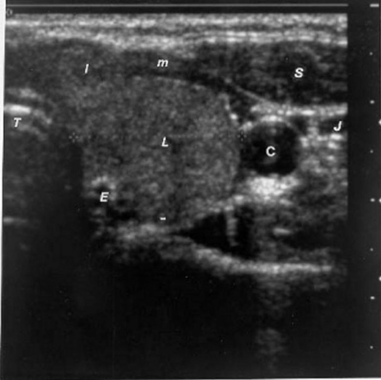
Ultrasonography Of The Thyroid - Thyroid Disease ManagerThyroid Disease
 www.thyroidmanager.org
thyroid anatomy ultrasonography sonography strap left sonogram gland goiter neck lobe muscles muscle enlarged carotid transverse blood radiology artery trachea
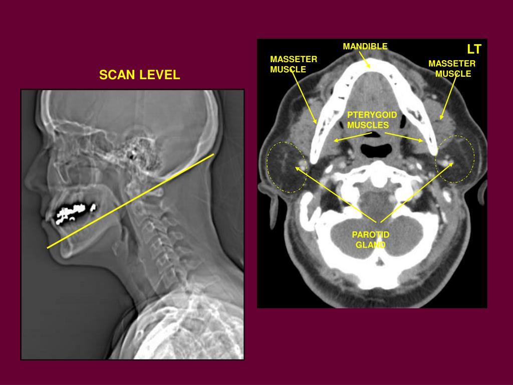
PPT - M-1 RADIOLOGY Head And Neck PowerPoint Presentation, Free
 www.slideserve.com
neck muscle lt radiology head masseter mandible ppt powerpoint presentation muscles pterygoid parotid gland scan level
Mylohyoid Muscle
 en.academic.ru
mylohyoid musculi colli
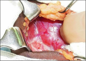
Anatomical Steps Of Thyroidectomy - Enlarged Thyroid
 khalidalomari.weebly.com
thyroid thyroidectomy steps right lobe medial enlarged
Soft Tissue Tumors Of The Head And Neck: Imaging-based Review Of The
 pubs.rsna.org
neck tumors sternocleidomastoid
Art Left Side Of The Neck, Transverse, Level II, CCDS. Two Lymph Nodes
 www.pinterest.com
lymph neck nodes level left ii node side muscle radiology sternocleidomastoid normal internal carotid artery transverse ccds fig pattern
Proliferative Myositis: A Rare Pseudomalignant Tumor Of The Head And
 jamanetwork.com
myositis proliferative tumor
Lymph neck nodes level left ii node side muscle radiology sternocleidomastoid normal internal carotid artery transverse ccds fig pattern. Ultrasonography of the thyroid. Sternocleidomastoid tumour in neonate: fibromatosis colli