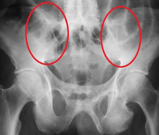
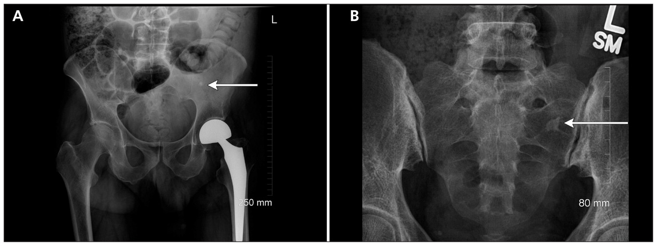
sclerosis of iliac bone
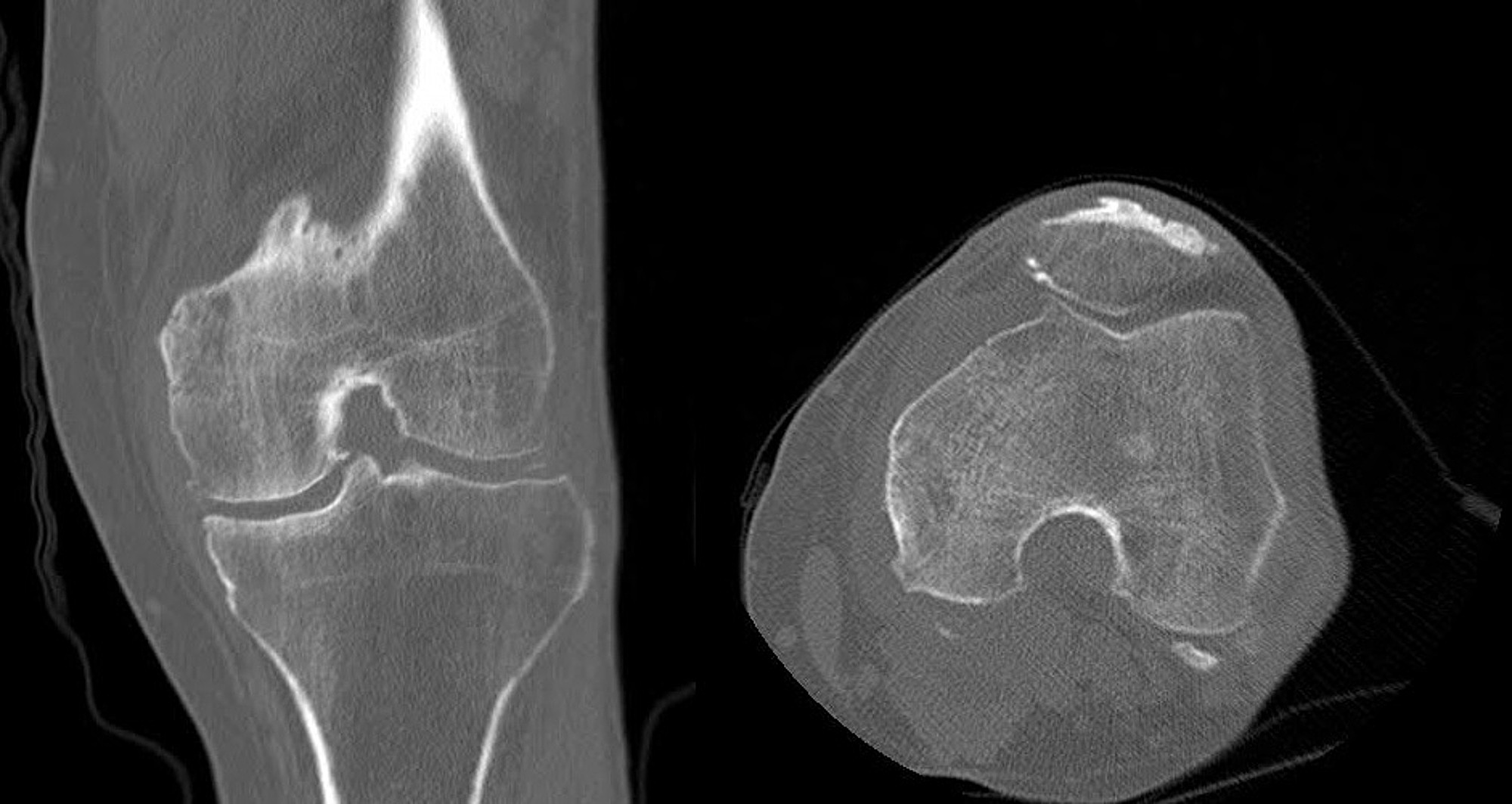
Fibrosarcoma Development 15 Years After Curettage and Bone Grafting of we have 9 Images about Fibrosarcoma Development 15 Years After Curettage and Bone Grafting of like Benign spotted bones: a diagnostic dilemma | CMAJ, Bone Island | Radiology Key and also Benign spotted bones: a diagnostic dilemma | CMAJ. Here it is:
Fibrosarcoma Development 15 Years After Curettage And Bone Grafting Of
 www.healio.com
fibrosarcoma bone lesion tumor cell giant femur distal lytic expansile cortex grafting curettage development years radiographs involving anteroposterior lateral showing
Benign Spotted Bones: A Diagnostic Dilemma | CMAJ
 www.cmaj.ca
cmaj benign dilemma
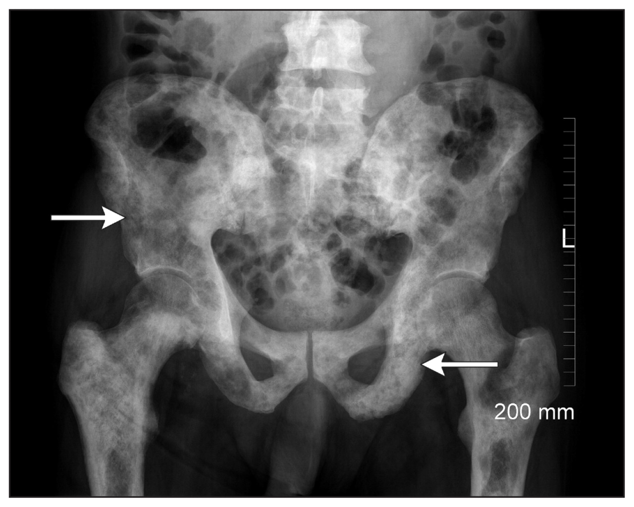
SACROILIAC JOINT DYSFUNCTION
 ptalert.blogspot.com
joint sacroiliac dysfunction si ankylosing joints fused widening changes ct sclerotic erosive bony
ScienceOpen
 scienceopen.com
cervical dislocation anterior spine syndesmophytes c4 lateral ray scienceopen c6 c5 ankylosis open joints c2 levels c3 figure
Benign Spotted Bones: A Diagnostic Dilemma | CMAJ
 www.cmaj.ca
benign cmaj dilemma powerpoint
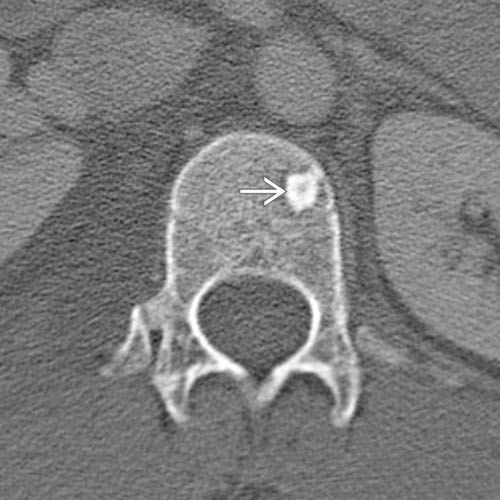
Bone Island | Radiology Key
 radiologykey.com
bone island ct sclerotic spine vertebral radiology body thoracic lesion sclerosis axial right dense margins focal reveals radiologykey
Cureus | An Unusual MRI Appearance Of Osseous Metastases
 www.cureus.com
mri bone ct window appearance metastases axial figure osseous unusual cureus coronal
Benign Spotted Bones: A Diagnostic Dilemma | CMAJ
 www.cmaj.ca
cmaj benign dilemma diagnostic
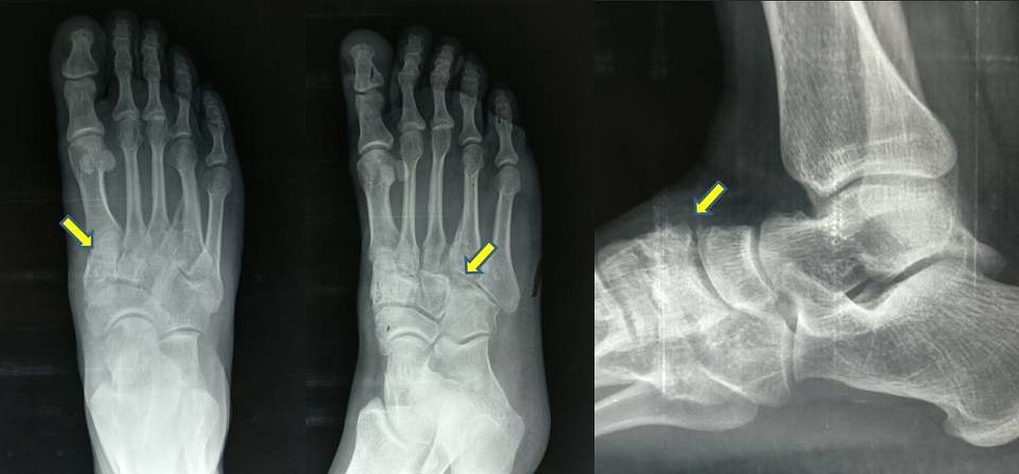
Cureus | Post-traumatic Arthritis Of The Tarsometatarsal Joint Complex
 www.cureus.com
joint tarsometatarsal arthritis traumatic lateral tmt joints second figure complex case report cureus radiograph posterior antero op pre views fourth
Bone island ct sclerotic spine vertebral radiology body thoracic lesion sclerosis axial right dense margins focal reveals radiologykey. Fibrosarcoma bone lesion tumor cell giant femur distal lytic expansile cortex grafting curettage development years radiographs involving anteroposterior lateral showing. Joint tarsometatarsal arthritis traumatic lateral tmt joints second figure complex case report cureus radiograph posterior antero op pre views fourth